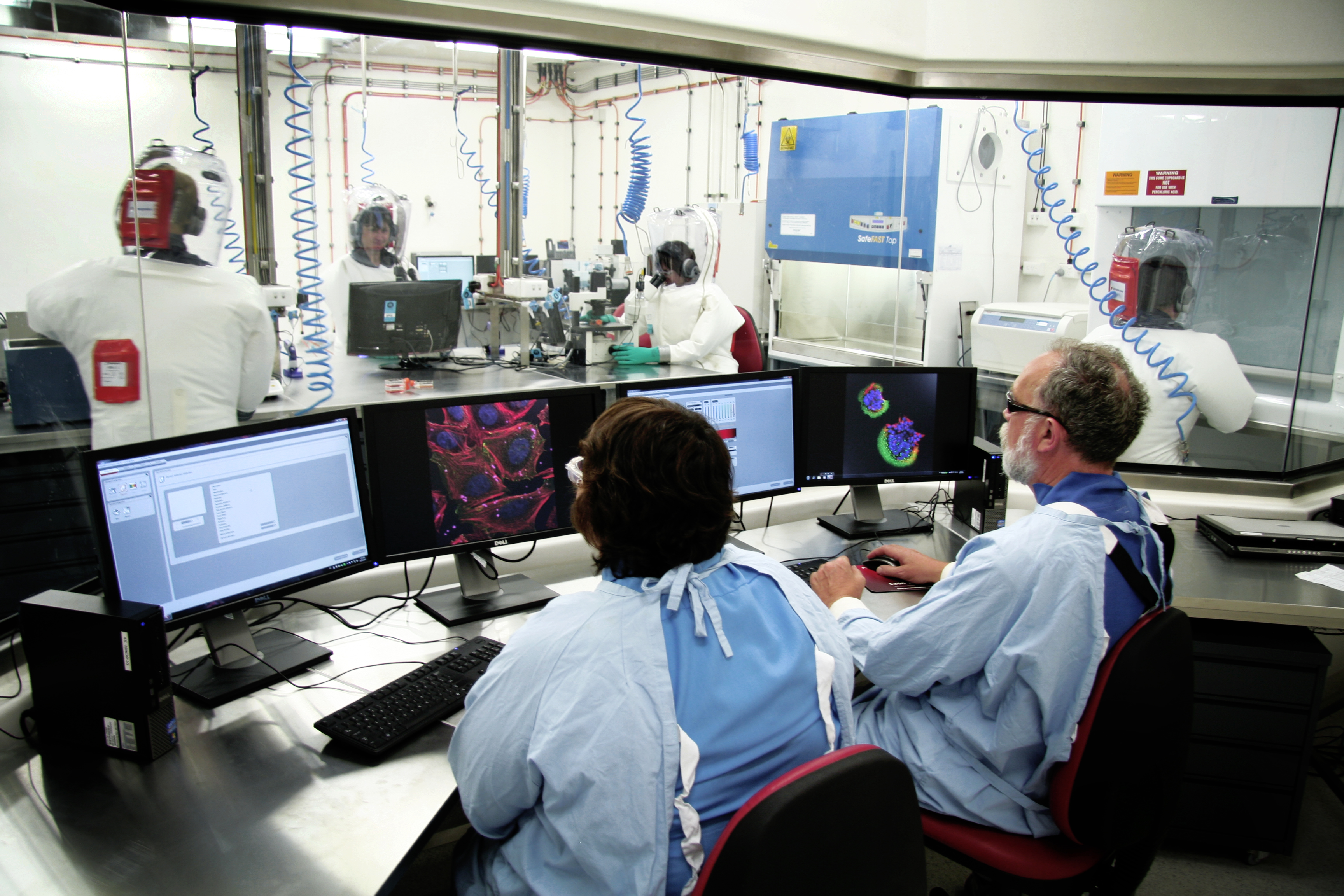
The Physical Containment (PC) 4 Zoonosis Suite and Bioimaging Facility is a shared resource dedicated to research into infectious diseases that affect the health of humans, domestic animals and wildlife. The Suite provides advanced technology and infrastructure for scientists undertaking research that requires a laboratory environment with the highest levels of biosafety.

Work performed here includes diagnostics, research specialising in the identification and characterisation of new and emerging viruses, comparative immunology and pathogenesis studies.
The PC4 Zoonosis Suite includes:
- two large laboratories (capable of operating at PC3 or PC4)
- specialist bioimaging equipment within the PC3/4 environment
- a dedicated PC3/PC4 training area
- insectary room at PC3/4
- two small animal rooms at PC3/4
Officially opened in December 2011, the Suite was constructed thanks to a grant from the National Collaborative Research Infrastructure Strategy (NCRIS), under the Networked Biosecurity Framework capability.
Bioimaging facility
Incorporated within the PC4 Zoonosis Suite, the Bioimaging Facility is a specialist microscopy service and a Linked Laboratory of the Australian Microscopy & Microanalysis Research Facility (AMMRF).
ACDP’s Bioimaging Facility is networked throughout Australia providing a real-time interactive platform for research collaboration.
Available instrumentation includes:
- Leica Laser micro-dissection LMD6500
- Leica confocal microscope SP5
- Olympus Live Cell light microscope
- JEOL 1400 Tomography transmission electron microscope
- Leica high pressure freezer HPM100.
Using specialised techniques, including viral identification, viral morphogenesis, advanced light microscopy, advanced transmission electron microscopy and scanning electron microscopy, our scientists have helped discover and characterise new viruses including:
- Hendra virus (previously known as equine morbillivirus)
- Herpesvirus of pilchards
- Epizootic haematopoietic necrosis virus (EHNV) of fish
- Aquabirnavirus (Tasmania)
- Nipah virus (Malaysia)
- Menangle virus
- Australian bat lyssavirus
- Bohle iridovirus
- Batrachochrytrium dendrobatidis (chytrid fungus).
Our equipment and expertise is available to research partners and includes:
- live cell imaging
- laser dissection microscopy
- confocal microscopy
- advanced transmission and scanning electron microscopy, including cryo immuno-electron microscopy, x-ray microanalysis and tomography.
Travel grants
Funding is available under the AMMRF Travel and Access Program (TAP) for short duration visits to ACDP and the Bioimaging Facility. The aim of this program is to provide access for researchers to state-of-the-art instrumentation and expertise.
There are three components to the TAP grants:
- a contribution towards airfare between the home city of the user and Geelong
- a contribution towards accommodation costs
- a contribution to the costs involved with instrument access.
The Physical Containment (PC) 4 Zoonosis Suite and Bioimaging Facility is a shared resource dedicated to research into infectious diseases that affect the health of humans, domestic animals and wildlife. The Suite provides advanced technology and infrastructure for scientists undertaking research that requires a laboratory environment with the highest levels of biosafety.
Work performed here includes diagnostics, research specialising in the identification and characterisation of new and emerging viruses, comparative immunology and pathogenesis studies.
The PC4 Zoonosis Suite includes:
- two large laboratories (capable of operating at PC3 or PC4)
- specialist bioimaging equipment within the PC3/4 environment
- a dedicated PC3/PC4 training area
- insectary room at PC3/4
- two small animal rooms at PC3/4
Officially opened in December 2011, the Suite was constructed thanks to a grant from the National Collaborative Research Infrastructure Strategy (NCRIS), under the Networked Biosecurity Framework capability.
Bioimaging facility
Incorporated within the PC4 Zoonosis Suite, the Bioimaging Facility is a specialist microscopy service and a Linked Laboratory of the Australian Microscopy & Microanalysis Research Facility (AMMRF).
ACDP’s Bioimaging Facility is networked throughout Australia providing a real-time interactive platform for research collaboration.
Available instrumentation includes:
- Leica Laser micro-dissection LMD6500
- Leica confocal microscope SP5
- Olympus Live Cell light microscope
- JEOL 1400 Tomography transmission electron microscope
- Leica high pressure freezer HPM100.
Using specialised techniques, including viral identification, viral morphogenesis, advanced light microscopy, advanced transmission electron microscopy and scanning electron microscopy, our scientists have helped discover and characterise new viruses including:
- Hendra virus (previously known as equine morbillivirus)
- Herpesvirus of pilchards
- Epizootic haematopoietic necrosis virus (EHNV) of fish
- Aquabirnavirus (Tasmania)
- Nipah virus (Malaysia)
- Menangle virus
- Australian bat lyssavirus
- Bohle iridovirus
- Batrachochrytrium dendrobatidis (chytrid fungus).
Our equipment and expertise is available to research partners and includes:
- live cell imaging
- laser dissection microscopy
- confocal microscopy
- advanced transmission and scanning electron microscopy, including cryo immuno-electron microscopy, x-ray microanalysis and tomography.
Travel grants
Funding is available under the AMMRF Travel and Access Program (TAP) for short duration visits to ACDP and the Bioimaging Facility. The aim of this program is to provide access for researchers to state-of-the-art instrumentation and expertise.
There are three components to the TAP grants:
- a contribution towards airfare between the home city of the user and Geelong
- a contribution towards accommodation costs
- a contribution to the costs involved with instrument access.
